
Types of imaging
CT
A CT (Computed Tomography) scan is carried out by a special type of x-ray machine. The images the machine produces are cross-sections of your body (think slices in a loaf of bread). The parts of your body can be shown in much greater detail than in standard x-ray films, and this helps the doctors diagnose your condition much more accurately.

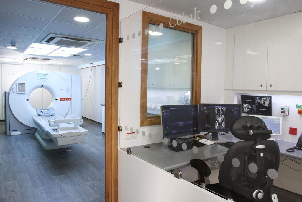
Imaging Technology
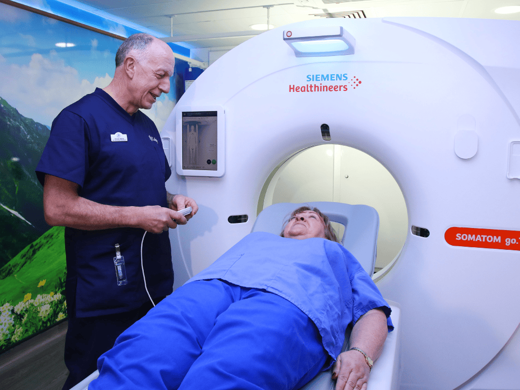
Siemens SOMATOM go.Top
Siemens SOMATOM go.All
What to Expect
When you arrive, please report to reception. Once you have been signed in, the radiographer will come and explain the procedure you are going to have. You should tell them if you have diabetes, asthma, or any allergies. If you need an oral contrast the radiographer will give you this before you go to the CT department. If you need to remove any clothing, someone will show you to the changing rooms and give you a patient gown or other special clothing to change into.
During the scan, you will be made comfortable on the moveable bed. Straps and pillows may be used to help you stay in the correct position and to help you stay still during the scan.
If you need an injection of contrast, you will be given this through a vein in your arm while you are lying on the scanner bed. This may make you feel warm and give you a metallic taste. The radiographer will stay with you during the injection. The radiographer will control
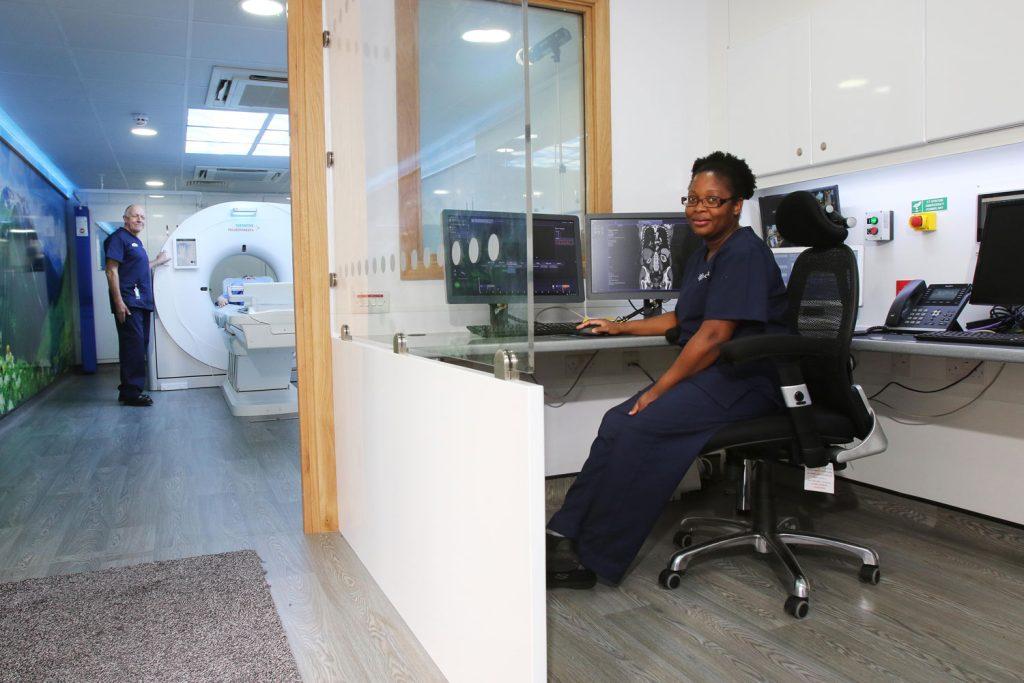
the bed from the control room and will slowly move it to position the part of your body being investigated inside the ‘doughnut.’
The radiographers will be in the control room during the scan, but you will be able to talk to them using an intercom, and they will be watching you all the time. You will hear a clicking and whirring sound from the CT scanner during the procedure. During the scan the radiographer may ask you to hold your breath or to not swallow while each image is being produced – if you feel any discomfort or anxiety because of this, please tell the radiographer immediately.
How long does a CT scan take?
Approximately 10 minutes.
The total time you will be in the department will be around an hour to an hour and a half.
How do I get my results?
Our specialist consultant will examine the CT images after your visit and will send a written report to your doctor which will normally be available within five to seven days. You will need to make an appointment with your doctor to discuss the results.

Referral FAQ's
Cobalt does not provide health screening scans. If a scan is clinically needed, we require a referral from a medical professional (this can be a consultant, a GP, a physiotherapist, a chiropractor, an osteopath, or an extended scope practitioner).
No, we do not accept self-referrals.
We need a medically qualified practitioner to refer you for a scan. This could be a Consultant, GP, Osteopath, Physiotherapist, Chiropractor, Extended Scope Practitioner or dentist. The medical practitioner cannot refer you for a scan outside their expertise and referrals are only accepted in line with their professional scope of practice. For example a musculoskeletal specialist can only refer you for a scan of your musculoskeletal system such as bones, joints and muscles
Reports are usually completed within a working week.
CT is computerised tomography and is used to provide a picture of the structure inside your body. A special type of X-ray machine is used to produce cross sectional images of your body. This involves being exposed to a small amount of radiation. The dose is equivalent to the amount of natural radiation you would receive over a period of three years.
MRI uses a powerful magnet providing high quality images. Structures in the body such as the brain, spine, breast, prostate, cartilage and ligaments in joints and muscles can be seen in much greater detail in all directions.